The human liver is needed for survival. This organ plays a very big role in metabolism, and other roles like glucose storage and most important, hormone production.The liver's specialized tissues are very important too
The urinary system (also called the excretory system) is the organ system that produces, stores, and eliminates urine. In humans it includes two kidneys, two ureters, the bladder and the urethra.
The nervous system is an organ system containing a network of specialized cells called neurons that coordinate the actions of an animal and transmit signals between different parts of its body. In most animals the nervous system consists of two parts, central and peripheral. The central nervous system of vertebrates (such as humans) contains the brain, spinal cord, and retina. The peripheral nervoussystem consists of sensory neurons, clusters of neurons called ganglia, and nerves connecting them to each other and to the central nervous system. These regions are all interconnected by means of complex neural pathways. The enteric nervous system, a subsystem of the peripheral nervous system, has the capacity, even when severed from the rest of the nervous system through its primary connection by the vagus nerve, to function independently in controlling the gastrointestinal system.
The muscular system is the anatomical system of a species that allows it to move. The muscular system in vertebrates is controlled through the nervous system, although some muscles (such as the cardiac muscle) can be completely autonomous
There are approximately 639 skeletal muscles in the human body.
The ear is the organ that detects sound. It not only receives sound, but also aids in balance and body position. The ear is part of the auditory system.
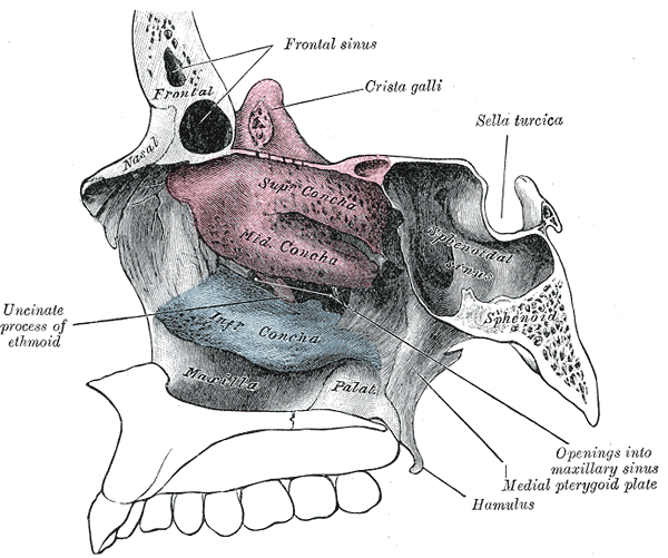
The nasal cavity conditions the air to be received by the other areas of the respiratory tract. Owing to the large surface area provided by the nasal conchae, the air passing through the nasal cavity is warmed or cooled to within 1 degree of body temparature. In addition, the air is humidified, and dust and other particulate matter is removed by vibrissae, short, thick hairs, present in the vestibule. The cilia of the respiratory epithelium move the particulate matter towards the pharynx where it passes into the esophagus and is digested in the stomach.
The diagram of the lateral wall of nasal cavity is shown below.
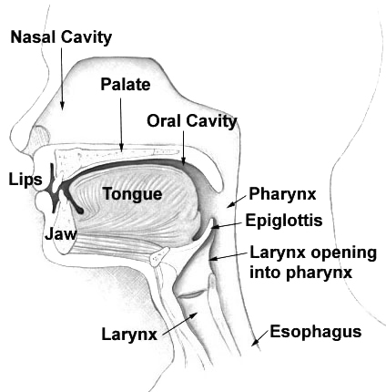
The esophagus (or oesophagus) is an organ in vertebrates which consists of a muscular tube through which food passes from the pharynx to the stomach. During swallowing, food passes from the mouth through the pharynx into the esophagus and travels via peristalsis to the stomach. The word esophagus is derived from the Latin œsophagus, which derives from the Greek word oisophagos , lit. "entrance for eating." In humans the esophagus is continuous with the laryngeal part of the pharynx at the level of the C6 vertebra. The esophagus passes through posterior mediastinum in thorax and enters abdomen through a hole in the diaphragm at the level of the tenth thoracic vertebrae. It is usually about 25–30 cm long depending on individual height. It is divided into cervical, thoracic and abdominal parts. Due to the inferior pharyngeal constrictor muscle, the entry to the esophagus opens only when swallowing or vomiting.
Let's take a look at the diagram of the oesophagus (esophagus).